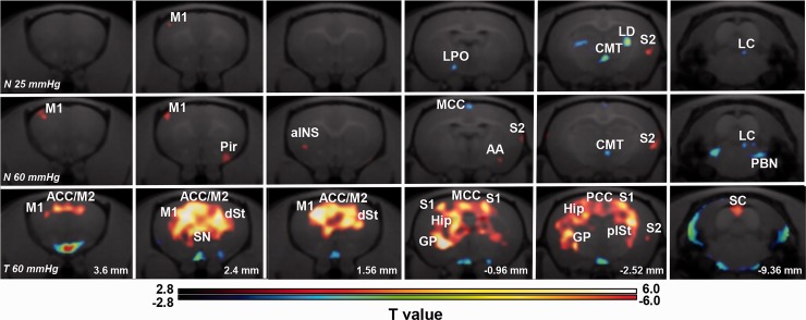
Figure 3.
Brain activity in response to different colorectal distension (CRD) pressures in normal and 2,4,6-trinitrobenzene sulfonic acid (TNBS)-injected rats. Upper line (N 25 mmHg): images showing activated (red) and suppressed (blue) brain regions in normal rats that received innocuous CRD with distention pressure at 25 mmHg (n = 9). Middle line (N 60 mmHg): images showing activated (red) and suppressed (blue) brain regions in normal rats that received noxious CRD with distention pressure at 60 mmHg (n = 11). Lower line (T 60 mmHg): images showing activated (red) and suppressed (blue) brain regions in TNBS-injected rats that received innocuous CRD with distention pressure at 60 mmHg (n = 13). Images from each group were obtained by voxel-based statistical comparison of FDG uptake with that in the normal group with CRD pressure at 0 mmHg. The T value of 2.82 was used as the threshold corresponding to the p < 0.005 (uncorrected) threshold. Right sides of images indicate the right hemisphere. Numbers in the lower line indicate the level of coronal slices in rat brain atlas (Paxinos and Watson). M1/M2: primary/secondary motor cortex; S1/S2: primary/secondary somatosensory cortex; Pir: piriform cortex; AA: anterior amygdaloid area; ACC: anterior cingulate cortex; MCC: midcingulate cortex; PCC: posterior cingulate cortex; aINS: anterior insular cortex; LPO: lateral preoptic area; dSt/plSt: dorsal/posterior lateral striatum; Hip: hippocampus; CMT: central medial thalamic nucleus; LD: laterodorsal thalamus nucleus; PBN: parabrachial nucleus; LC: locus coeruleus; SC: superior colliculus; GP: globus pallidus; SN: septal nucleus.

