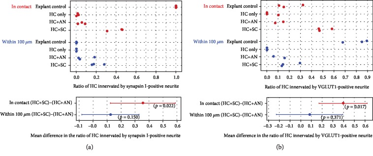
Figure 2.
Quantification of stem cell-derived innervation of HCs within 12 days of coculture. The graphs depict the ratio of HCs innervated by synapsin I-positive neurites (a) and VGLUT1-positive neurites (b) in both control conditions and with SCs. The ratio is calculated as the observed innervation of HCs divided by the total numbers of HCs in the preparation. Innervation is shown in red (dots) and close proximity (within 100 μm) is shown in blue (dots). Quantification of innervation for within the 100 μm group did not include those puncta in contact with the HC. The ratio of HCs innervated by synapsin-positive neurites (a) and VGLUT1-positive neurites (b) was significantly higher (p < 0.05, independent samples t-test) in HC-SC cocultures compared with HC-AN cocultures. HC: hair cell; AN: auditory neurons; SC: differentiated stem cells.

