Figure 2.
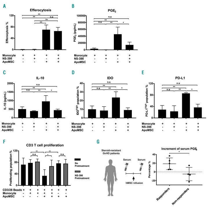
Efferocytosis-induced COX2/PGE2 is the key effector molecule of immunosuppressive monocytes. (A) NS-398 (100 μM) was added during the co-culture of monocytes and apoptotic mesenchymal stromal cells (ApoMSC). After 8 h, efferocytosis was evaluated using flow cytometry, n=3. As reported in (A), PGE2, n=4 (B) and IL-10, n=4 (C) were evaluated in cell culture supernatants using enzyme-linked immunosorbent assays, while IDO, n=5 (D) and PD-L1, n=3 (E) in monocytes were examined by flow-cytometry. (F) COX2 activity in efferocytosing monocytes was inhibited by using 100 μM NS-398 before adding them to CellTrace™ Violet-labeled CD3 T cells. Proliferation of T cells was measured and analyzed by flow cytometry, n=4. Experimental data are expressed as means ± standard deviation. One-way analysis of variance and the post-hoc Tukey test were used to compare the mean differences among the samples. (G) Eight steroid-resistant patients with graft-versus-host disease (GvHD) receiving mesenchymal stromal cells (MSC) were analyzed for serum PGE2 levels. The percentages of PGE2 increment [PGE2after infusion - PGE2before infusion]/PGE2before infusion × 100%] were compared between clinical responders (● n=5) and non-responders (■ n=4). An unpaired t test was used to compare the mean differences between two groups (*P<0.05; **P<0.01; ns: not significant).

