Figure 5.
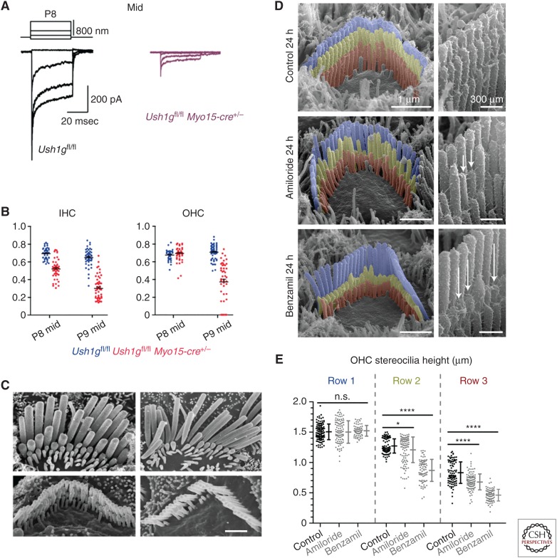
Tip links and transduction regulate stereocilia length. (A) Mechanoelectrical transduction (MET) currents recorded from outer hair cells (OHCs) located in the middle of the cochlea in Ush1gfl/fl mice (black traces, left) and Ush1gfl/fl Myo15-cre+/− mice (right, pink) mice at P8. (B) Height of stereocilia in row 2 (R2) relative to that in row 1 (R1) in inner hair cells (IHCs) (left) and OHCs (right) in the middle of the cochlea in Ush1gfl/fl mice (blue) and Ush1gfl/fl Myo15-cre+/− mice (red) mice at P8 and P9. The second row stereocilia are shorter in the IHCs of Ush1gfl/fl Myo15-cre+/− mice at P8 and continue to shorten by P9. The second row stereocilia of the OHCs of Ush1gfl/fl Myo15-cre+/− mice start to shorten at P9. (C) Scanning electron micrographs showing the hair bundles of IHCs (top) and OHCs (bottom) in the middle of the cochlea at P9 (left) and at P22 (right). Note the shortening and loss of the second and third row stereocilia. Scale bar, 1 μm. (A–C from Caberlotto et al. 2011; adapted, with permission, from the authors in conjunction with National Academy of Sciences © 2011 Open Access policy.) (D) Scanning electron micrographs showing hair bundle from early postnatal mice (P4-P6) that were cultured in control medium (top), 100 µM amiloride (middle), and 30 µM benzamil (bottom) for 24 hours. Stereocilia have been pseudocolored; row 1 (blue), row 2 (yellow), row 3 (red). MET channel block by amiloride and benzamil cause a rapid shortening of the second and third rows. (E) Quantification of data shown in D above. Significant shortening is observed in rows 2 and 3 but not in row 1. n.s., Not significant. *p < 0.05, ****p < 0.001. (D and E from Vélez-Ortega et al. 2017; adapted under the terms of the Creative Commons Attribution License.)

