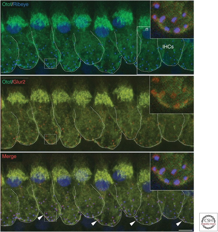
Figure 5.
Colocalization between presynaptic ribbons and postsynaptic GluA2 in mature inner hair cells (IHCs). A confocal z-stack projection of IHCs triple-immunostained for otoferlin (green), the ribbon protein RIBEYE (blue), and the GluA2 subunit of postsynaptic AMPA receptors for glutamate (red). At mature IHC synapses, all RIBEYE and GluA2 puncta “co-localize” (arrowheads indicate just a few of them). Dotted lines provide a rough indication of the basolateral membrane around the IHC synaptic region. Note that the RIBEYE antibody also stained the IHC nuclei (n). The insets (top-right corners) are an expanded view of the IHC synaptic region. Scale bar, 5 µm.

