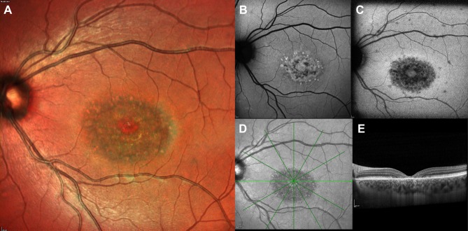
Figure 1.
Multimodal imaging of a patient with STGD1 and macular involvement. Color fundus photograph (A), near-infrared-autofluorescence (B) and short wavelength fundus autofluorescence (C). Absence of RPE is visible as an area of irregular hyopoautofluorescence on fundus autofluorescence. Several hyperautofluorescence lesions at the macula correspond to the flecks. The OCT scan (D) centered on the fovea shows atrophy of RPE and ellipsoid zone and backscattering.

