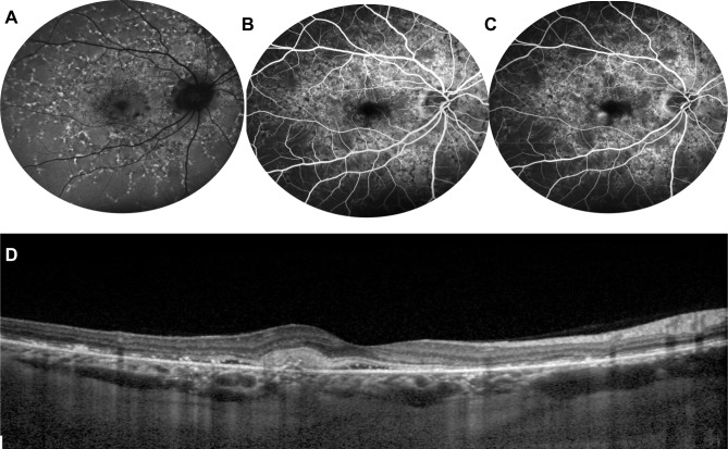
Figure 6.
Choroidal neovascularization in STGD1. Fundus autofluorescence (A) shows hyperautofluorescent flecks s and irregular macular hypoautofluorescence. Early (B) and late (C) fluorescein angiography frames demonstrate a parafoveal focal dye leakage, corresponding to a type 1 choroidal neovascularization (CNV). OCT scan (D) reveals growth of the CNV above the RPE with mild exudation, along with focal irregularity/atrophy of RPE/ellipsoid zone layers.

