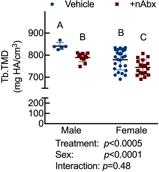
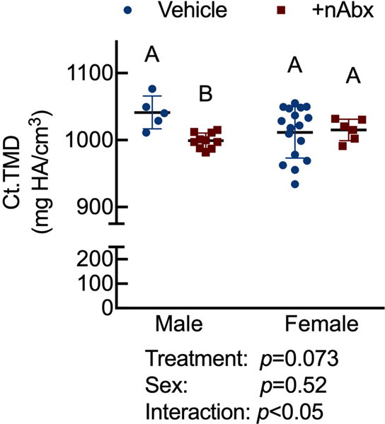
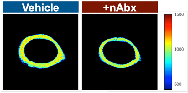
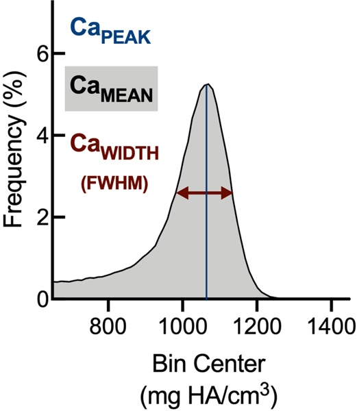
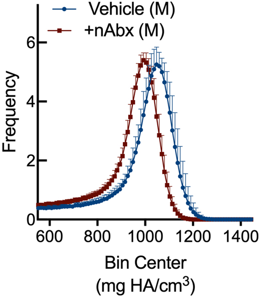
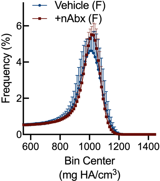
Figure 4. Influence of neonatal Abx on bone tissue material properties.
(A) Tissue mineral density in distal femur secondary spongiosa; n=5–11 male or 18–26 female mice per condition.
(B) Tissue mineral density in midshaft femur; n=5–10 male or 6–17 female mice per condition.
(C) Hydroxyapatite-calibrated heatmap of tissue mineral density distribution in mid-diaphyseal cortical bone from male and female mice gavaged with water or neonatal Abx.
(D) Histograms of mid-diaphyseal TMDD quantification. (i) Diagrammatic representation of CaPEAK, CaMEAN, and CaWIDTH; TMDD in (ii) male and (iii) female mice.
Columns represent mean ± standard deviation; groups with different letters are statistically different from each other.

