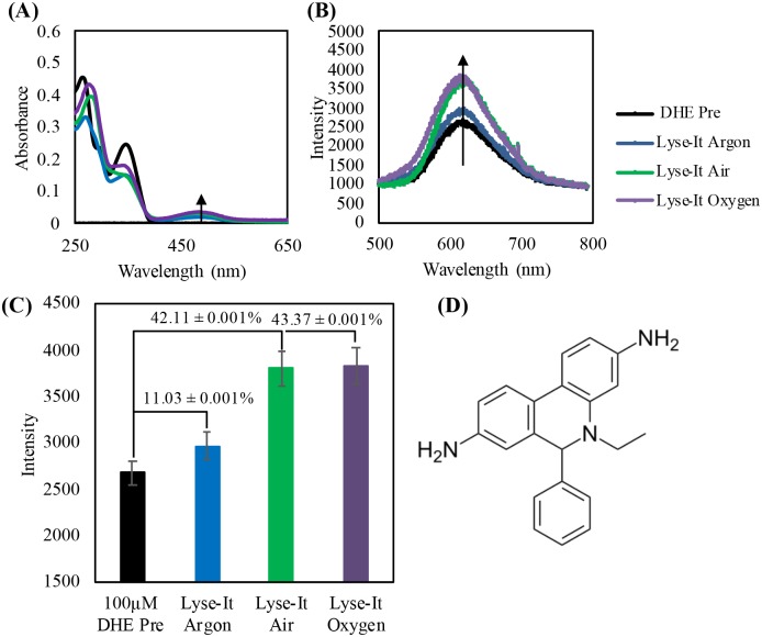
Fig 6.
100 μM DHE purged then irradiated at 50% power, 60 seconds with Lyse-It (A) Absorption spectra of DHE pre and post microwaved irradiation, (B) Fluorescence spectra pre and post purging and microwave irradiation, (C) Fluorescence λmax intensity with the percentage increase from Pre with respect to increasing oxygen concentration. (D) Dihydroethidium unreacted probe. As oxygen content increases, the peak at approximately 475-nm in the absorbance spectra and 605-nm in the fluorescence spectra increases indicating an increase in the detection of superoxide anion radical.

