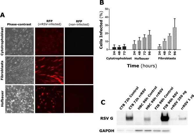
Fig 1. RSV infection of primary human placental cells.
(A) Isolated human placental cells (left panels) after 72 hours (cytotrophoblast cells) or 96 hours (fibroblast and Hofbauer cells) infection at MOI of 1.0 with an RSV-A2 strain (rrRSV) expressing red fluorescent protein (RFP). Strong RFP expression resulting from rrRSV replication was observed in fibroblast and Hofbauer, but not in cytotrophoblast cells (center panels). Matched non-infected controls did not exhibit any fluorescence (right panels). (B) Proportion of cytotrophoblast, fibroblast, and Hofbauer cells exhibiting red fluorescence as a result of rrRSV replication. Each time-point represents the mean ± SEM of 8 different fields. (C) Expression of RSV G protein in cytotrophoblast (CTB), Hofbauer (HBC), and fibroblast (FIB) cells infected with rrRSV and processed at 72 or 96 hours for Western blot analysis. rrRSV lysate was used in two different concentrations (250 ng and 1 μg) as a positive control.

