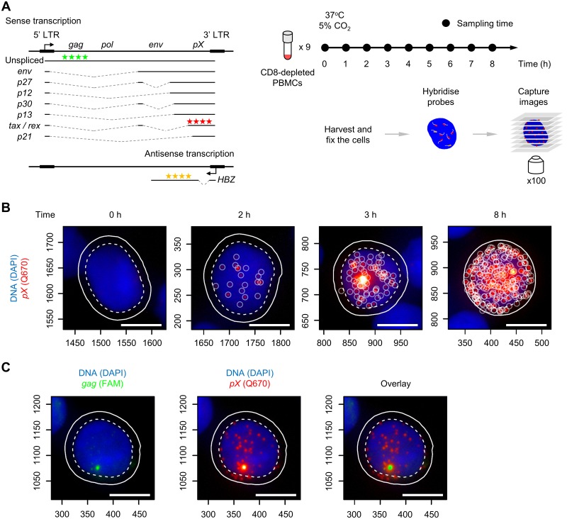
Fig 1. Intense transcription of the HTLV-1 sense strand in PBMCs identified by smFISH.
(A) HTLV-1 transcripts, smFISH probes and sample preparation for smFISH. Three sets of probes for smFISH are indicated that hybridize to HTLV-1 transcripts: sense-strand in the pX region (Q670, red stars), HBZ (Q570, yellow stars), and unspliced sense transcripts containing gag region (FAM, green stars). (B) Representative images of HTLV-1+ PBMCs with the sense-strand transcripts at the indicated time points. Blue area indicates the nucleus stained with DAPI, and red spots indicate the HTLV-1 sense transcripts. Scale bar (white) = 5 μm. (C) A site of intense transcription identified by the probes for gag. The image on the left shows the gag staining (green). The image overlaid with sense transcript spots (red, shown in the middle) is presented on the right. The cell presented is from a HAM patient coded TDZ sampled at 4 hours of incubation.

