
Figure 5. The Mus musculus domesticus Gut Microbiome Confers a Survival Advantage after Lethal IAV Infection.
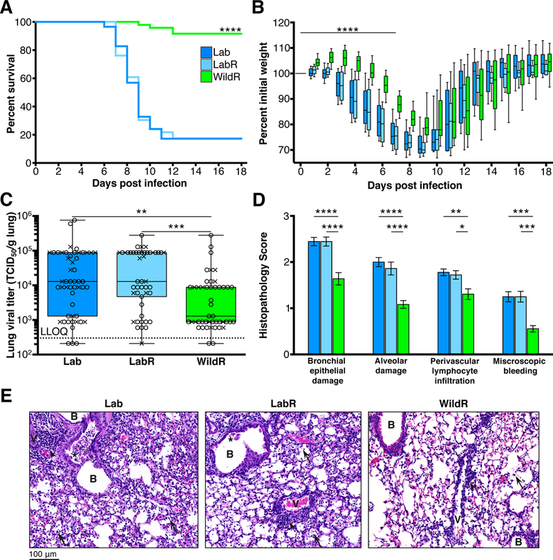
Female mice were inoculated with 400 TCID50 and male mice with 600 TCID50 of PR8. (A and B) Mice were monitored daily for 18 days and mice that lost 30% or more of their body weight were euthanized (Lab,n=58; LabR, n=46; WildR, n=48). (A) Kaplan Meier survival curves, ****P<0.0001 comparing WildR with either LabR or Lab by log rank (Mantel-Cox) analysis. (B) Weight loss curves, ****P<0.0001 comparing the slope of the weight loss (day 0 to day 7) of WildR to that of LabR or Lab in a linear regression analysis. Weight loss of LabR did not significantly differ from that of Lab. Median and IQR are presented. (C) Lung viral titer 3 days post IAV infection assessed via MDCK monolayers in 96-well plates using an antibody-based assay. Lab, n=51; LabR, n=49; WildR, n=48. Median and IQR are presented. (D) Histopathological scores 7 days post IAV infection. B: Bronchi; V: Vessels; arrows: lymphocytes and/or red blood cells in alveoli; arrowheads: perivascular lymphocyte infiltration; asterisks: bronchial epithelial cell death. Mean and SEM are presented. (E) Representative lung histology 7 days post IAV infection (original magnification 40x). Lab, n=18; LabR, n=18; WildR, n=18. *P<0.05, **P<0.01, ***P<0.001, ****P<0.0001. Significance was determined using parametric One-Way ANOVA with Tukey multiple comparison test with 95% confidence interval (Gaussian model), or Kruskal-Wallis with Dunn’s multiple comparison test. All data shown are from three independent experiments using both female (X) and male (O) mice.See also Figure S7.