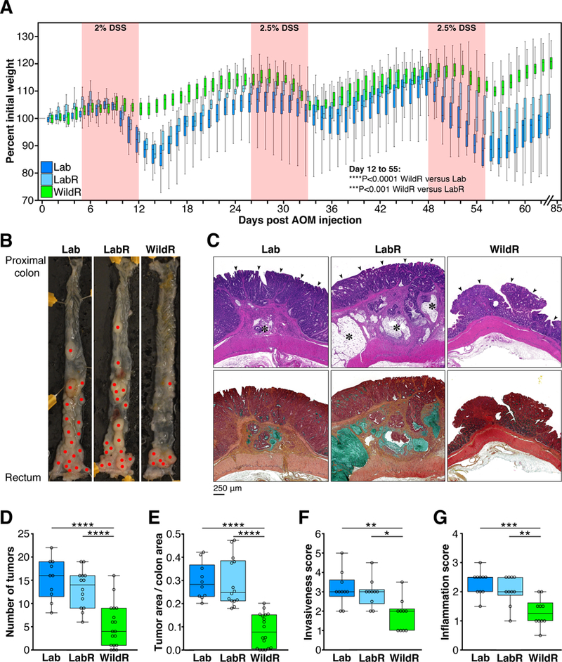
Figure 7. The Mus musculus domesticus Gut Microbiome Confers Protection from Colitis-Associated Tumorigenesis.

Clinical data from male mice that received a single intraperitoneal injection of AOM (10 mg/kg of body weight), followed by three 7 day cycles of 2–2.5% DSS in drinking water. Mice were monitored for weight loss throughout the time-course of the experiment and euthanized on day 85 to assess tumor burden. (A) Weight loss curves following AOM/DSS treatment. Lab, n=18; LabR, n=16; WildR, n=19. WildR vs. Lab, ****P<0.0001, and WildR vs. LabR, ***P<0.001 (repeated measures mixed model linear regression). Weight loss of LabR did not significantly differ from that of Lab. (B) Representative images of dissected colons (red dots indicate tumors). (C) Representative colon histology (original magnification 10x). Upper panel: HandE staining of longitudinal colon tumor sections; arrowheads: well differentiated adenocarcinoma in mucosa. LabR and Lab tumors invade the submucosa and muscular layers with moderate to severe inflammation; asterisks: mucinous nodules. Lower panel: Movat’s staining of serial sections of the same tumors as in the upper row images. Mucinous nodules (mucin stained in green) containing mucinous carcinoma cells and tubular adenocarcinoma lining are found in submucosa and muscle layers of LabR and Lab tumors. Lab, n=10; LabR, n=10; WildR, n=10. (D) Number of tumors. (E) Fraction of total colon area covered in tumors. Tumor burden was assessed using ImageJ software. (F) Invasiveness scores based on tumor location. (G) Inflammation score based on inflammatory cell infiltration. Median and IQR are presented. *P<0.5, **P<0.01, ***P<0.001, ****P<0.0001. Significance was determined using parametric One-Way ANOVA with Tukey multiple comparison test with 95% confidence interval (Gaussian model). All data shown are from three independent experiments.
