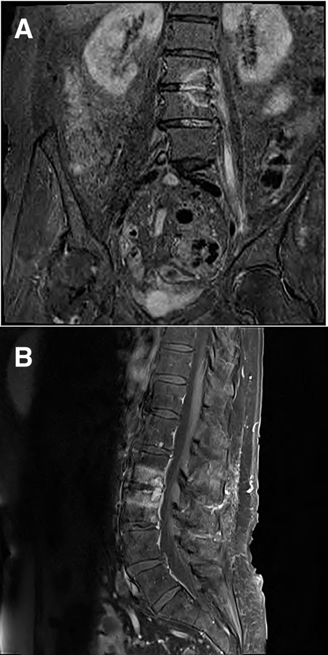
Figure 1.

(A) MRI T2 fat suppressed coronal image of the lumbar spine showing L2–L3 high signal abnormality with involvement of the left psoas muscle in keeping with acute spondylodiscitis and psoas abscess formation. (B) MRI T1 fat suppressed sagittal image of the lumbar spine showing L2–L3 high signal abnormality with involvement of the left psoas muscle in keeping with acute spondylodiscitis and psoas abscess formation.
