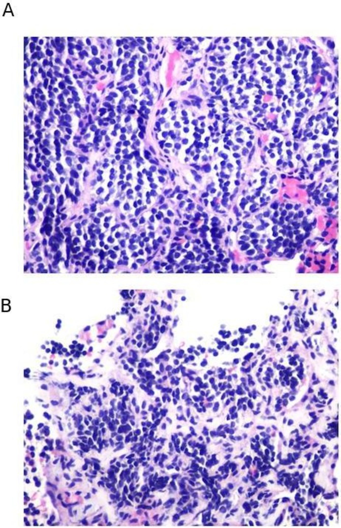
Figure 2.

Pathology of core biopsies. Analysis of core biopsies of supraclavicular lymph node (A) and abdominal subcutaneous tissue (B) were significant for metastatic neuroendocrine tumour. These biopsies were performed prior to treatment with nivolumab and ipilimumab. Lymph node tissue was hypercellular with syncytial group and numerous single-lying hyperchromatic cells with coarse chromatin (A). Abdominal subcutaneous tissue was hypercellular composed of loosely cohesive groups and single-lying cells with high N/C ratio, containing finely granular chromatin pattern. No necrosis or mitoses were identified (B).
