
Fig. 4.
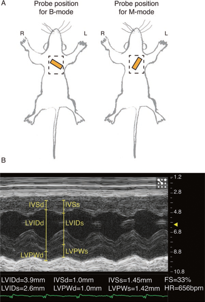
Echocardiography recording and analysis. (A) Approximate positioning of echocardiography probe (orange rectangle) for a 2D long-axis B-mode view (left) and short-axis M-mode view (right). Dashed rectangle indicates the recommended area from which to remove hair. (B) Parasternal short-axis M-mode with left ventricular functional measurements. Post capture, left ventricle internal dimension in diastole/systole (LVIDd/s); left ventricle posterior wall thickness in diastole/systole (LVPWd/s); interventricular septum thickness in diatole/systole (IVSd/s); fractional shortening (FS); and heart rate (HR) are measured