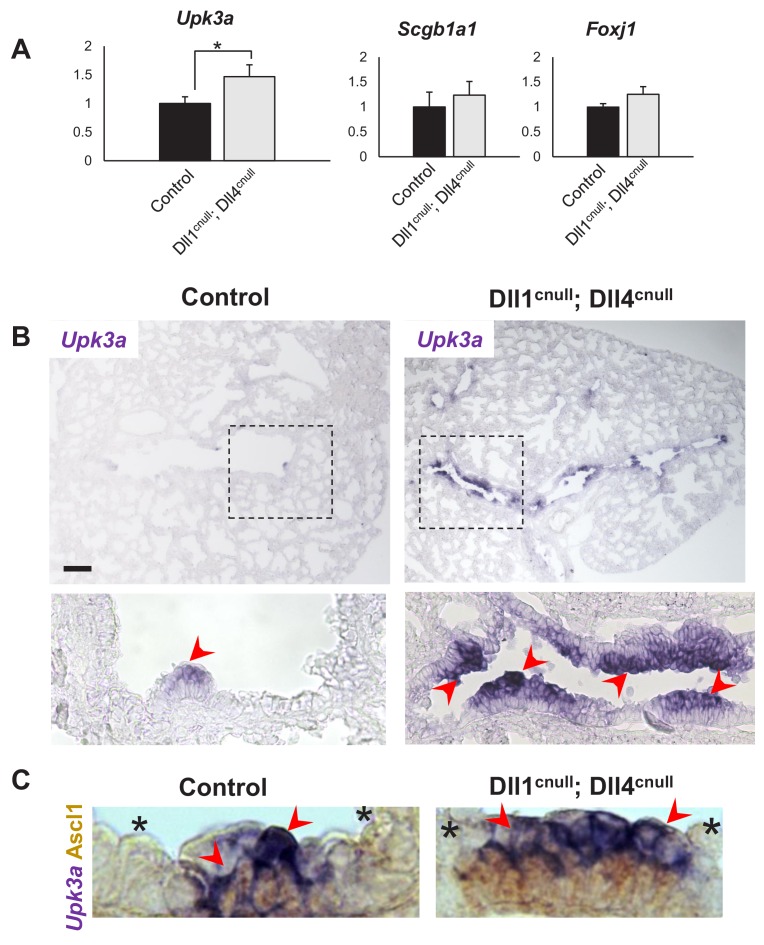
Figure 6. Expansion of the Upk3a expression domain in Dll1cnull; Dll4cnull lungs.
(A) qPCR analysis: significantly increased expression of Upk3a, but not of Scgb1a1 or Foxj1 in mutants relative to controls (n = 3 in both groups). Graphs are mean ± SEM. Student’s t-test *p<0.05. (B) ISH for Upk3a in E18.5 lungs showing marked expansion of the Upk3a expression domain (arrowheads) in intrapulmonary airways of Dll1cnull; Dll4cnull mutants (boxed areas enlarged in the lower panels). (C) Double immunohistochemistry (Ascl1)/ISH (Upk3a) confirms that the Upk3a+ cells (arrowheads) are NEB-associated CCs. Note reciprocal high (arrowhead) and low (*) intensity of signals in areas outside and inside the NEB microenvironment, respectively. Scale bar in B = 40 μm.

