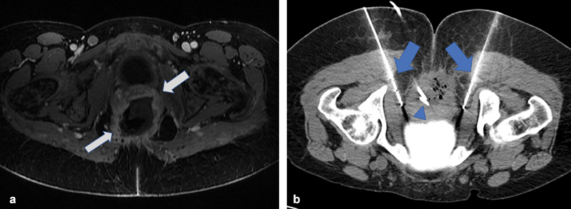
Fig. 3.

( a ) Axial T1-weighted fat-saturated post-contrast MRI image in the pelvis demonstrates a large pelvic mass (arrows) with a necrotic center in a patient with known advanced cervical carcinoma hospitalized for intractable pain. ( b ) Axial intraprocedural image demonstrates placement of two cryoablation probes (arrows) adjacent to the proximal pudendal nerves. Incidental note is made of a percutaneously placed drainage catheter (arrowhead).
