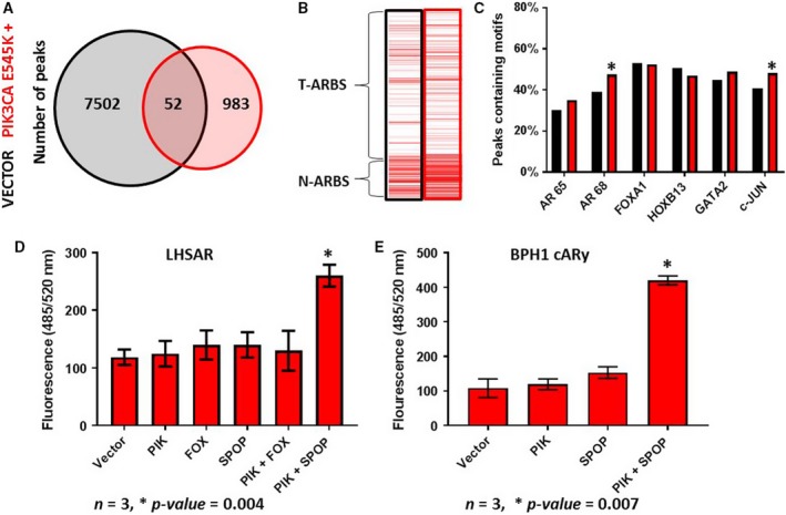
Figure 4.

Mutations that cause the AMS are insufficient to transform cells on their own. The benign LHSAR prostate cells were transfected with a PIK3CA E545K expression plasmid or control, and AR ChIP‐seq was performed 2 days later. A Venn diagram of shared and unique peaks compared to controls (A) demonstrates a reduction of ARBSs. Enrichment for Pomerantz T‐ARBSs and N‐ARBSs was performed (B) and shows increased enrichment of N‐ARBSs. The percentage of TF motifs was determined (C) and suggests that PIK3CA mutant did not induce the AMS. (D) The indicated expression plasmids were transfected into LHSAR cells alone or in combinations, and a soft agar colony‐forming assay was performed. Growth was only observed with the combination of the PIK3CA and SPOP mutations. (E) The experimental results were recapitulated in the benign BPH1‐AR cells, which express ectopic AR. Statistical analysis of motif enrichment between groups was by chi‐square test using r software environment; a two‐way t‐test was used for analysis of the colony‐forming assay; a P‐value of > 0.05 was considered significant as indicated by a *; and error bars are SEM where n = 3.
