
Figure 1.
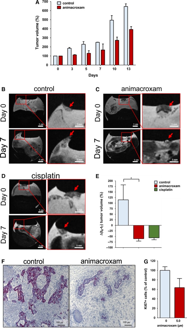
Antineoplastic effects of animacroxam and cisplatin in vivo. (A) Relative tumor volume of TGCT xenografts grown in athymic nude mice after the treatment with 0.9% NaCl or animacroxam (daily dose of 60 mg·kg−1 body weight on four consecutive days, intraperitoneal). Results are shown as mean ± SEM of n = 2 mice in each group. (B–D) T2w MR images of tumor‐bearing CAMs of fertilized chicken eggs taken before and after 7 days of a single intravenous injection with 0.9% NaCl (B), 5.0 µm animacroxam (C), or 2.5 µm cisplatin (D); scale bar = 1 cm (left side) or 0.5 cm (right side). (E) Mean changes in tumor volume of n = 3 CAM experiments. (F) IHC staining of Ki67 (red–violet) revealed a pronounced reduction of proliferation in animacroxam‐treated tumors as compared to untreated controls; scale bar = 100 µm. (G) Quantification of Ki67‐positive cells as a marker for proliferation in tumors excised out of the CAM after incubation with animacroxam (5.0 µm). Results are shown as mean ± SEM of n = 4 independent preparations. *P‐values of ≤ 0.05, unpaired t‐test.