Figure 6.

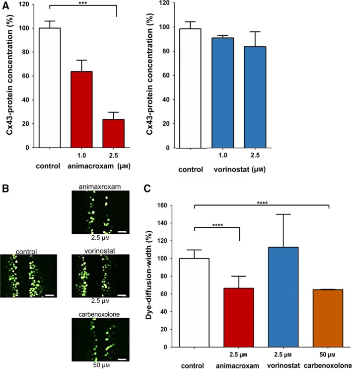
Treatment‐induced changes in Cx43 expression and endothelial cell–cell communication. (A) Protein expression of Cx43 was reduced after 24 h of incubation with animacroxam (1.0–2.5 µm) or—albeit to a lesser extent—with vorinostat (1.0–2.5 µm) when compared to control (100%). (B) Representative images of Lucifer Yellow dye diffusion in endothelial EA.hy926 cells after incubation with animacroxam or vorinostat; scale bar = 100 µm. (C) Lucifer Yellow dye diffusion, indicating intercellular gap‐junctional cell–cell coupling, was diminished after 24 h of incubation with animacroxam (2.5 µm). This effect was absent after incubation with vorinostat (2.5 µm). The established Cx43 inhibitor Cbx (50 µm) served as a positive control. Results are shown as means ± SEM of at least n = 3 independent experiments. ***P‐values of ≤ 0.0005, ****P‐values of ≤ 0.0001; one‐way ANOVA post hoc Tukey’s test.
