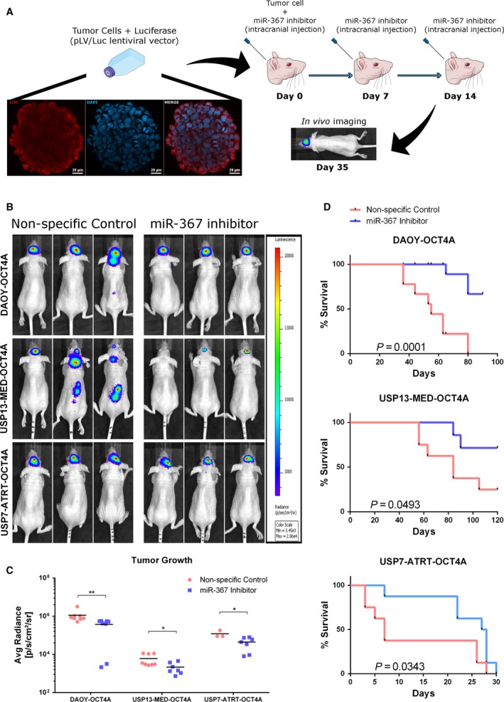
Figure 5.

Therapeutic targeting of miR‐367 in mice bearing orthotopic OCT4A‐overexpressing embryonal CNS tumor xenografts. (A) Schematic overview of the in vivo experimental layout. A suspension of 106 tumor cells was injected into the right lateral ventricle of Balb/C nude mice at day 0, and series of oligonucleotides were injected at days 0, 7, and 14. Representative immunofluorescence images of Daoy tumorsphere incubated with antibody against firefly luciferase (red) and DAPI (blue). Scale bar: 20 μm. (B) Representative bioluminescence‐based images of OCT4A‐overexpressing tumors in mice, 35 days post‐intracerebroventricular cell injection. (C) Bioluminescence intensity analysis of USP7‐ATRT, USP13‐MED, and DAOY tumors at day 35. Data are expressed as mean ± SEM (*P < 0.05, **P < 0.01, t test compared with respective control condition). (D) Overall survival rates of mice bearing OCT4A‐overexpressing embryonal CNS tumors (log‐rank Mantel–Cox test; n = 8 per group).
