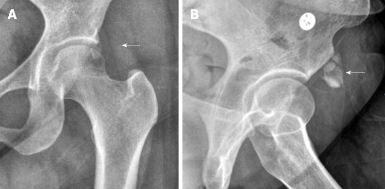
Figure 1.
An anteroposterior radiography of the left hip. A: An anteroposterior radiography of the left hip showing an amorphous calcification (white arrow) adjacent to the anterior inferior iliac spine which is the attachment site of the rectus tendon, suggesting calcific tendinopathy of rectus femoris; B: The lesion (white arrow) was more clearly defined at the lateral view.

