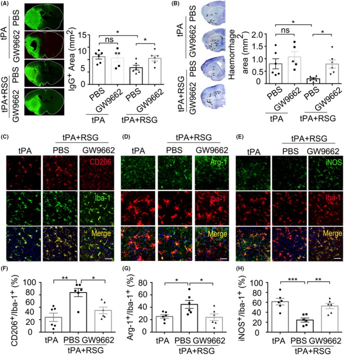
Figure 7.

PPAR‐γ antagonist GW9662 abolished the protection of RSG against HT and BBB disruption in tPA‐infused stroke mice. A, Endogenous IgG extravasation into the parenchyma was visualized by staining sections with antibodies against mouse IgG molecules. B, Representative examples of images of intracerebral hemorrhage identified on cresyl violet (CV)‐stained coronal section. C, Representative confocal images of CD206 and Iba‐1 double immunostaining in the brains obtained from tPA‐infused stroke mice 1 day after MCAO treated with tPA, RSG and RSG + GW9662. Scale bar = 100μm. D, Representative confocal images of Arg‐1 and Iba‐1 double immunostaining in the brains obtained from tPA‐infused stroke mice 1 day after MCAO treated with tPA, RSG and RSG + GW9662. Scale bar = 50μm. E, iNOS and Iba‐1 double immunostaining 1 day after MCAO in mice treated with tPA, tPA + RSG and tPA + RSG+GW9662. Scale bar = 100μm. F, Quantification of the percentage of CD206+/Iba‐1 + cells in the brain, n = 6 per group. G, Quantification of the percentage of Arg‐1+/Iba‐1 + cells in the brain, n = 6 per group. H, Quantification of the percentage of iNOS+/Iba‐1 + cells in the brain, n = 6 per group. Data are expressed as mean ± SEM. * P ≤ .05, ** P ≤ .01 vs tPA. PPAR‐γ, Peroxisome proliferator‐activated receptor‐γ; RSG, rosiglitazone; tPA, tissue plasminogen activator; HT, hemorrhagic transformation; MCAO, middle cerebral artery occlusion; iNOS, inducible nitric oxide synthase; Arg‐1, arginase 1; Iba‐1, ionized calcium‐binding adaptor molecule 1
