Figure 1.

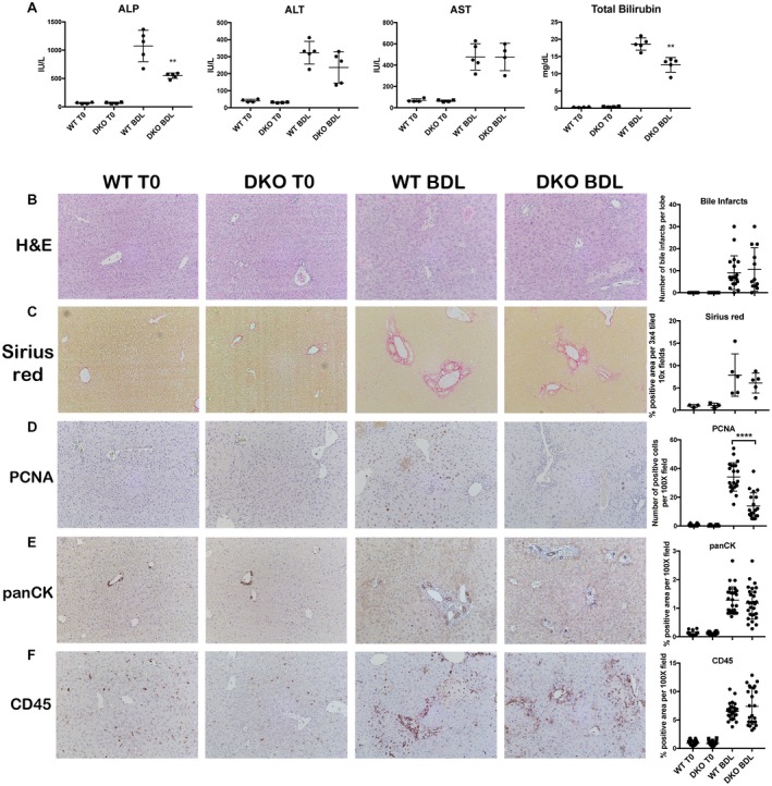
LRP5/6 KO (DKO) mice have decreased biliary injury but equivalent parenchymal injury, fibrosis, inflammation, and ductular response after BDL. (A) Both ALP and bilirubin are significantly decreased in DKO livers at 14 days after BDL compared to WT livers, while AST and ALT are equivalent; **P < 0.01 versus WT+BDL (t test). (B) No change in the number of bile infarcts in WT and DKO mice after BDL (magnification ×100). (C) Sirius red staining shows equivalent fibrosis in DKO mice after BDL compared to WT mice (magnification ×100). (D) IHC shows fewer PCNA + proliferating hepatocytes in DKO mice after BDL compared to WT mice (magnification ×100); ****P < 0.0001 versus WT+BDL (t test). (E) IHC for panCK shows that ductular response is unchanged between DKO and WT mice after BDL (magnification ×100). (F) Inflammation is equivalent in DKO and WT mice after BDL, as assessed by CD45 IHC (magnification ×100). Horizontal lines in the graphs represent mean ± SEM. Abbreviations: H&E, hematoxylin and eosin; IU, international units; PCNA, proliferating cell nuclear antigen.
