Figure 2.

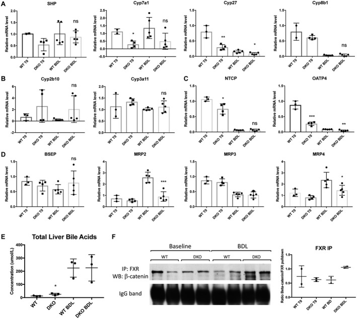
Hepatic BAs in DKO mice are comparable to WT mice after BDL despite changes in BA metabolism genes. (A) Expression of SHP is unchanged between WT and DKO mice, while Cyp7a1 and Cyp27 are decreased in DKO mice before and after BDL. (B) Detoxification enzymes Cyp2b10 and Cyp3a11 are not induced in DKO mice after BDL. (C) Expression of uptake transporter OATP4 is decreased in DKO mice before and after BDL, and NTCP is decreased at baseline. (D) Expression of efflux transporter MRP2 is decreased in DKO mice after BDL, while BSEP is unaffected; additionally, MRP4 is significantly decreased in DKO mice compared to WT mice after BDL. (E) Total liver BAs are elevated in DKO mice at baseline but are equivalent to WT mice after BDL. (F) IP with FXR shows maintenance of the FXR/β‐catenin complex in DKO mice before and after BDL. For A‐E, *P < 0.05, **P < 0.01, and ***P < 0.001 versus WT (t test). Data in the graphs represent mean ± SEM. Abbreviations: IgG, immunoglobulin G; mRNA, messenger RNA; ns, not significant.
