Figure 3.

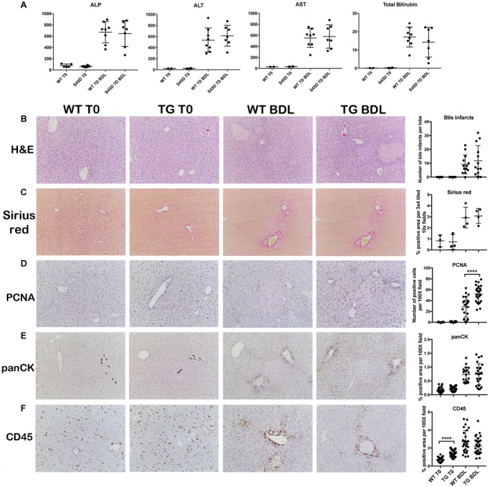
Overexpression of mutated nondegradable S45D β‐catenin does not increase injury, fibrosis, inflammation, or ductular response following BDL. (A) There is no difference in hepatic (AST, ALT) or biliary (ALP, bilirubin) injury between WT and TG mice after BDL. (B) No change in the number of bile infarcts in WT and TG mice after BDL (magnification ×100). (C) Sirius red staining shows equivalent fibrosis in TG mice after BDL compared to WT mice (magnification ×100). (D) IHC shows an increased number of PCNA+ proliferating hepatocytes in TG mice after BDL compared to WT mice (magnification ×100); ****P < 0.0001 versus WT+BDL (t test). (E) Ductular response is equivalent between TG and WT mice after BDL, as assessed by panCK IHC (magnification ×100). (F) Although CD45 staining is increased in TG compared to WT mice at baseline, there is no difference in inflammation between TG and WT mice after BDL (magnification ×100). Data in the graphs represent mean ± SEM. Abbreviations: H&E, hematoxylin and eosin; PCNA, proliferating cell nuclear antigen.
