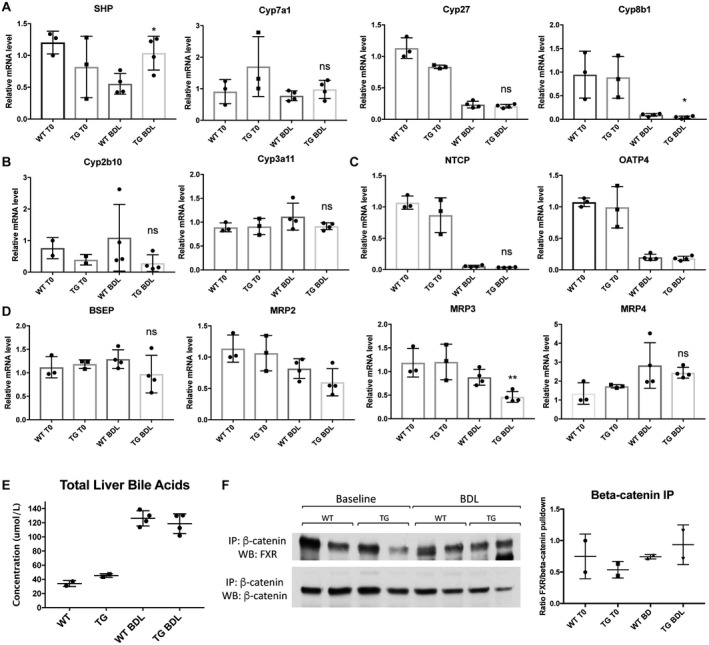
Figure 4.

BA metabolism and FXR/β‐catenin association are comparable in TG and WT mice after BDL. (A) Expression of SHP is increased in TG compared to WT mice after BDL, while Cyp7a1 and Cyp27 are unchanged before and after BDL. (B) Detoxification P450 enzymes are equivalently expressed in WT and TG mice after BDL. (C) BA uptake is unaffected by overexpression of β‐catenin after BDL. (D) Expression of apical and basolateral BA exporters are not different between WT and TG mice in response to BDL, with the exception of MRP3, which is decreased in TG compared to WT mice. (E) Total liver BAs are equivalent in TG and WT mice after BDL. (F) IP with β‐catenin shows maintenance of the FXR/β‐catenin complex in TG mice before and after BDL. For A‐E, *P < 0.05 and **P < 0.01 versus WT (t test). Data in the graphs represent mean ± SEM. Abbreviations: mRNA, messenger RNA; ns, not significant.
