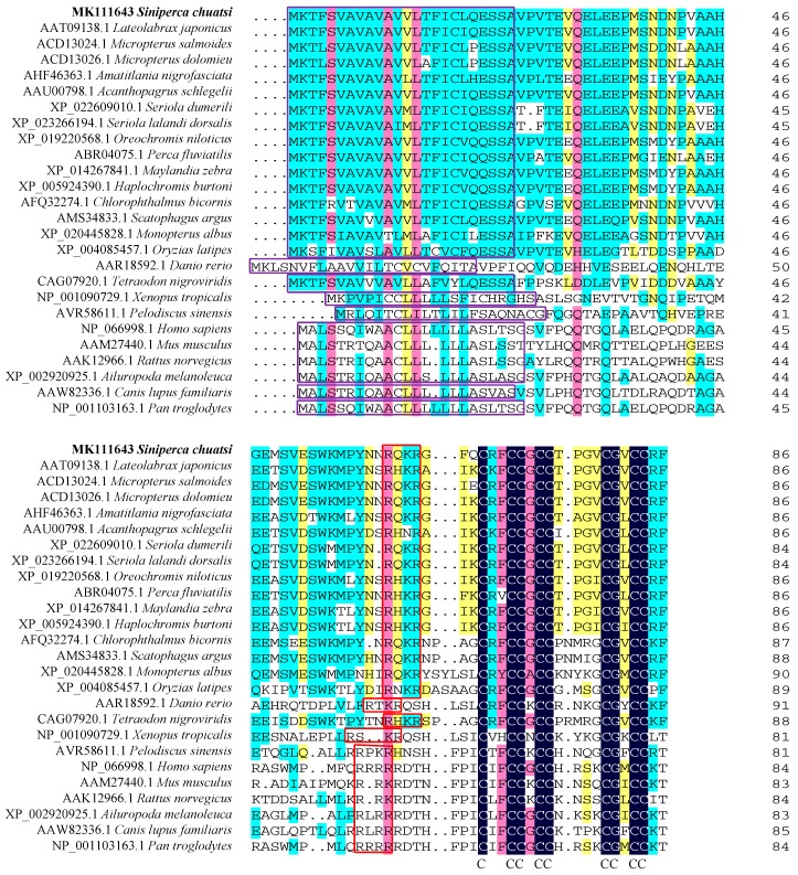
Figure 2.
Multiple-sequence alignment of the hepcidin protein. The dark blue regions indicate completely identical amino acid sequences. The pink, cyan, and yellow regions indicate amino acid similarities greater than 75%, 50%, and 33%, respectively. The purple and red squares indicate signal peptide and RX(K/R)R motif, respectively. Mazarine represents the conserved eight cysteine residues.

