Figure 5.
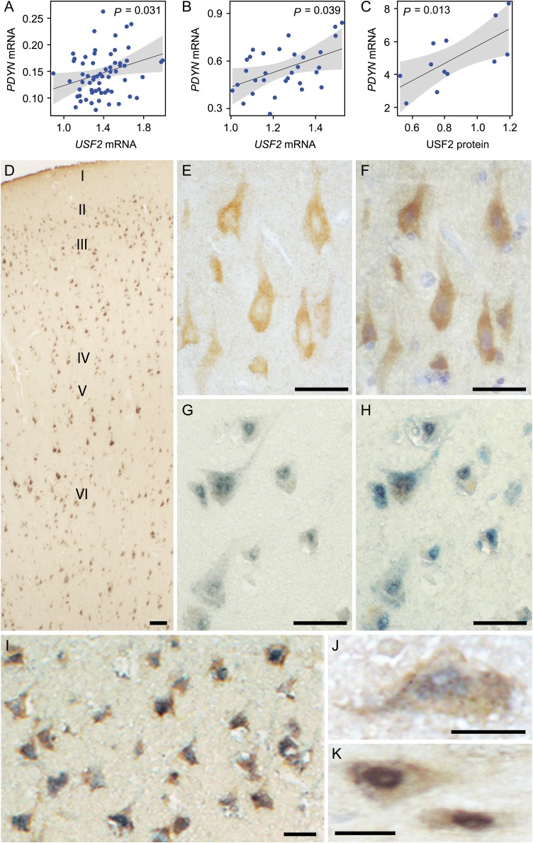
USF2 is positively associated with PDYN mRNA and is co-localized with PDYN in the human dlPFC. (A–C) Effect display for the main effect of USF2 mRNA (A, n = 64 subjects of the First cohort; B, n = 31 subjects of the Second cohort) and USF2 protein (C, n = 12 subjects) on PDYN mRNA. 95% confidence band is drawn around the estimated effect. P-values from ANOVAs are indicated. Potentially confounding factors such as age, postmortem interval, tissue pH, RBFOX3, GFAP levels, and alcoholism were included as covariates in the analysis but did not cause any significant effects. (D) Overview of dlPFC laminae immunolabeled for PDYN and USF2 proteins. Note the uniform distribution of the signal abundance and intensity across cortical laminae (shown with roman numerals). (E) A representative image showing PDYN immunoreactivity in neurons of the layer V. Note predominantly cytoplasmic localization of the signal. (F) Visualization of cell nuclei in (E) by hematoxylin staining. (G) A representative image showing USF2 signal in the layer V. Note predominantly nuclear localization of the signal. (H) Visualization of cell nuclei in (F) by toluidine blue staining. (I) Both PDYN and USF2 immunoreactivity are located in the same neurons in the layers III/IV. (J and K) High-magnification images of neurons in the layer V expressing both PDYN and USF2. Scale bars, 100 μm (D); 50 μm (E–I); and 25 μm (J and K).

