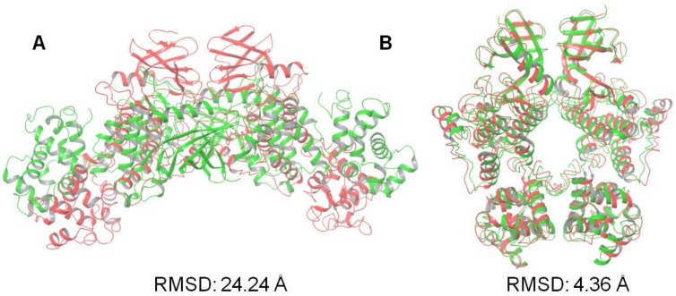
Figure 4.
Illustration of the protein–protein docking results of KIRA-containing IRE1 monomers (PDB code: 4U6R). Values shown are the RMSD in angstrom of the positions of the Cα atoms of the best-scoring docked pose (green) against the native IRE1 dimer structure in (A) face-to-face (PDB code: 3P23) and (B) back-to-back (PDB code: 4YZC) conformation (red).

