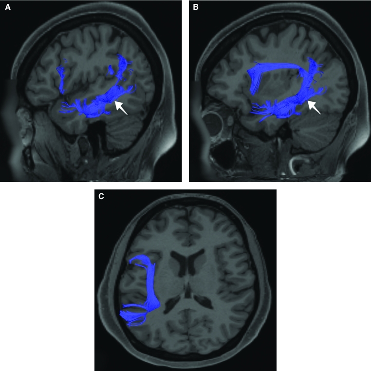
FIGURE 6.
Structural connectivity of STSdp in the left hemisphere, shown on T1-weighted MR images. Sagittal views of A, lateral and B, medial planes and C, axial views showing projections to the frontal and occipital lobes. Dark blue: white matter tracts of STSdp demonstrating connections with arcuate/SLF and the “u” fibers of the occipitotemporal system.

