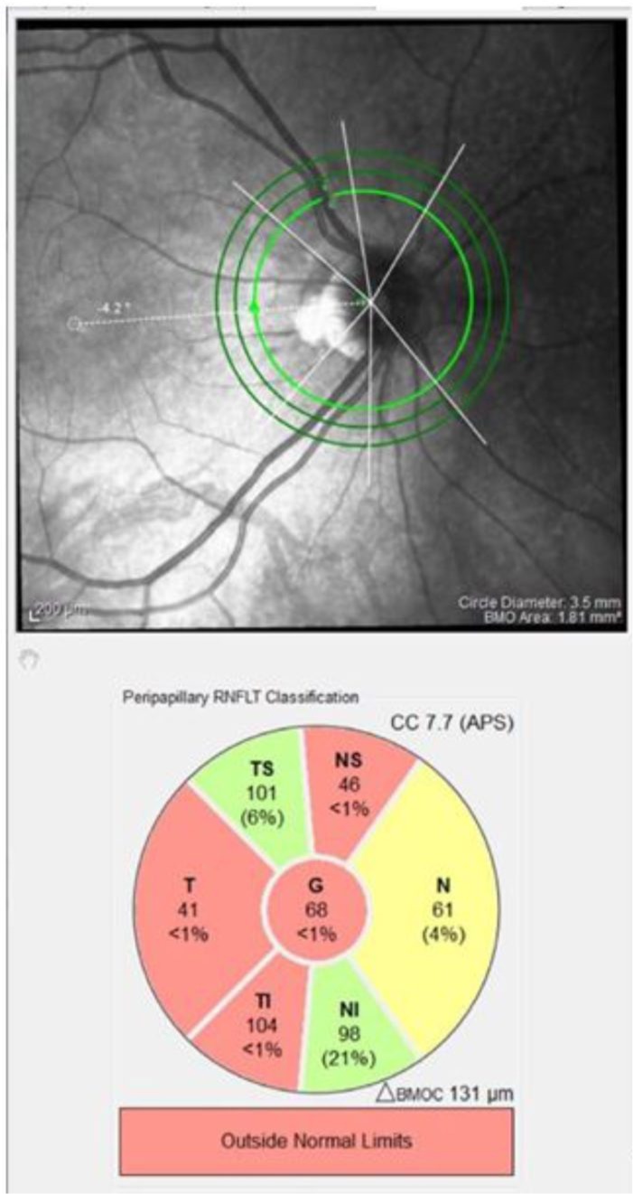
Figure 2.

An SD-OCT pRNFL scan of the right eye of a patient who has had ON, demonstrating temporal pRNFL thinning. Red sectors indicate those outside normal limits.
pRNFL, peripapillary retinal nerve fibre layer; ON, optic neuritis; SD-OCT, spectral domain optical coherence tomography.
