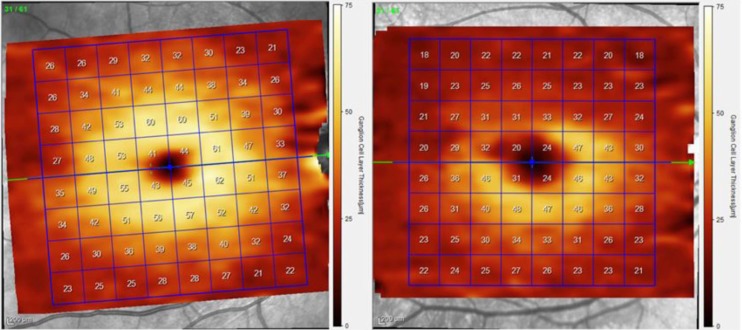
Figure 3.
Ganglion cell layer thickness maps from a segmented SD-OCT scan of the macula from the right eye of a healthy (left) and ON (right) patient. The segmented GCL is selected in this example, demonstrating superior loss of ganglion cells and corresponding thinning.
GCL, ganglion cell layer; ON, optic neuritis; SD-OCT, spectral domain optical coherence tomography.

