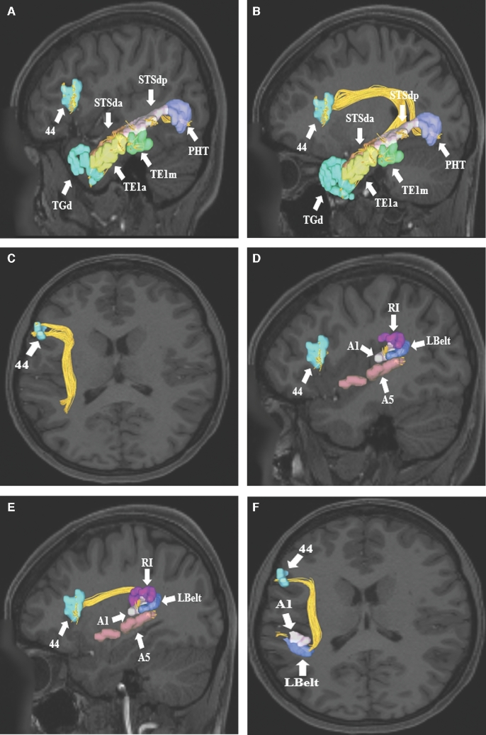
FIGURE 8.
A-C, SLF connections from area 44 in the inferior frontal gyrus to parts of temporal lobe. Connections are shown in the left cerebral hemisphere on T1-weighted MR images in the A and B, sagittal and C, axial planes. Area 44 exhibits structural connections via the SLF to temporal regions TGd, TE1a, TE1m, STSda, STSdp, and PHT in this subject brain. D-F, Area 44 also demonstrates structural connections to areas A5, A1, LBelt, and RI in the superior temporal gyrus and posterior insular cortex. Connections are shown in the left cerebral hemisphere on T1-weighted MR images in the D and E, sagittal, and F, axial planes. All parcellations are identified with white arrows and corresponding labels. The SLF can be seen coursing posteriorly before bending 90° to enter the subcortical white matter of the temporal lobe and terminate in the aforementioned parcellations.

