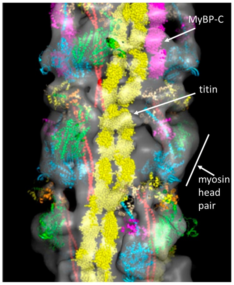
Figure 4.
3D reconstruction of part of the bridge region of myosin filaments from human cardiac muscle [20]. The image shows a length of about 450 Å, containing three crowns of head pairs. The possible location of strands of titin and the accessory protein C-protein (MyBP-C) are shown in yellow and mauve respectively. The myosin head densities have been fitted with interacting head motif structures ([21]; discussed later). This conformation is supposed to be the position of the heads in what has been called the super-relaxed state when the ATP turnover rate is very low (see Section 2.4). Adapted from [20].

