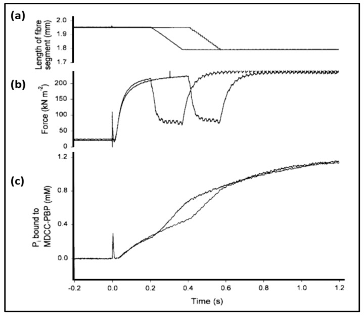
Figure 16.
Simultaneous measurement of length (a), force (b) and phosphate release (c) in a single skinned muscle fibre, illustrating the acceleration of Pi release rate during shortening. A permeabilized muscle fibre was mounted between two hooks, one attached to a length-adjusting motor and the other to a force transducer. The Figure shows two consecutive measurements on a single fibre. The fibre was initially at rest length in a rigor solution (no ATP). At zero time a contraction was initiated by the release into a muscle fibre of around 1.5 mM ATP by laser photolysis of caged ATP. (a) The length of the fibre as controlled by the motor. At either 0.2 or 0.4 s, the fibre was allowed to shorten by 8% of its length. (b) Force measurements: after approximately 0.2 s, the fibre, which initially was prevented from shortening, reached its maximum level of force development. (c) The amount of phosphate bound to the Pi-sensor incubated with the muscle fibre. The trace shows clearly that the rate of Pi liberation was increased during shortening steps (adapted from Figure 5 of He et al. [116]).

