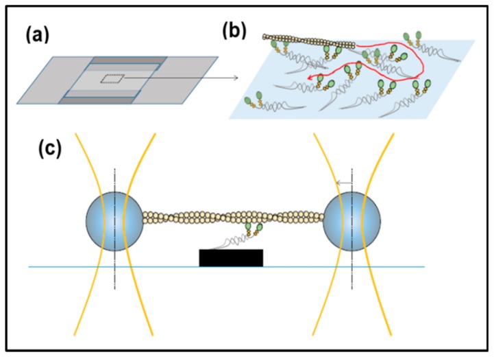
Figure 17.
In vitro motility assay and optical tweezers set-up for single-molecule mechanics. (a) Flow cell of two cover-slips sandwiched on top of each other via spacers; (b) magnified view of rectangular area in (a) indicating the principle for the gliding in vitro motility assay with surface-adsorbed myosin heads that propel an actin filament. The curved red arrow indicates a possible filament sliding path; (c) schematic diagram of the three-bead optical tweezers assay where an actin filament is suspended between two beads held in optical traps. The filament is then lowered down to allow the actin filament to interact with single myosin motor fragments adsorbed to a third bead or another type of pedestal as indicated here. Reproduced from Special Issue Review [125] with permission.

