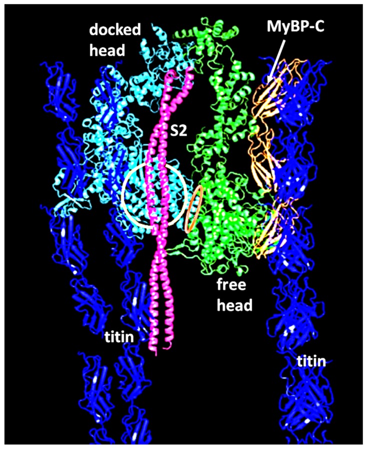
Figure 22.
Part of the IHM structure in Figure 4 [21] as detailed by Marston [144]. Strands of titin are shown in dark blue, three domains of MyBP-C in orange, the myosin subfragment-2 (S2) in pink, and the myosin heads in pale blue (docked head) and green (free head). The myosin mesa on the docked head is circled in orange and the free head converter domain interacting with the docked head is indicated by a red ellipse. Adapted from Figure 2c of Marston [144] using structural data from [20].

