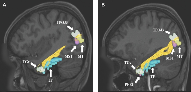
FIGURE 3.
A and B, ILF connections from temporal regions TGv, TF, and PEEC to the temporal-parietal-occipital junction (TPOJ3) and lateral occipital lobe (MT and MST). Connections are shown in the left cerebral hemisphere on T1-weighted MR images in the sagittal plane. All parcellations are indicated with white arrows and corresponding labels. The ILF is seen coursing from the temporal regions posteriorly to the occipital lobe.

