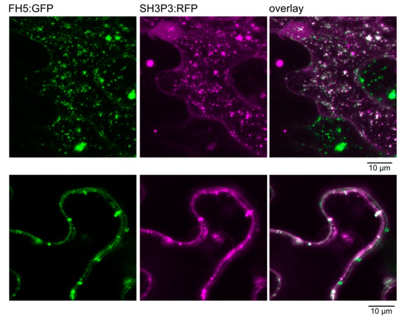
Figure 3.
Co-localization of FH5, C-terminally tagged by green fluorescent protein (GFP), with SH3P3, C-terminally tagged by red fluorescent protein (RFP), in transiently transformed N. benthamiana leaf epidermis, as documented by confocal laser scanning microscopy. Note the cell exhibiting only GFP fluorescence and the weak nuclear RFP signal, indicating absence of cross-channel signal leak. Top: Maximum intensity projection of a Z-stack of optical sections across an epidermal pavement cell. Bottom: A single confocal section showing a detail of an anticlinal cell to cell boundary.

