Figure 4.
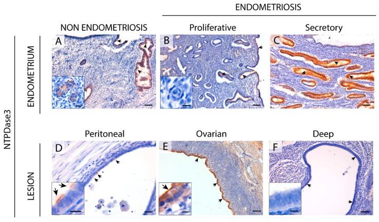
Immunolocalization of NTPDase3 in the eutopic (A–C) and ectopic (D–F) endometrial tissue. NTPDase3 was immunodetected in ciliated and non-ciliated cells of cyclic endometrium from women without (A) and those with (B,C) endometriosis (arrows), with changes in expression along the menstrual cycle, reaching a maximum at the secretory phase (C). Moreover, NTPDase3 was present in the endothelial cells of spiral arteries of women without endometriosis (inset in A) but not in the cyclic endometria from women with the disease (inset in B). In ectopic endometrial tissue, NTPDase3 was weakly expressed in the epithelial cells (arrows) of peritoneal lesions (D) and highly expressed in the epithelium (arrows) of the ovarian endometriomas (E). NTPDase3 was absent in the deep infiltrating lesions (F, vaginal nodule) (arrows). Insets in images (D–F) are details of the epithelium of the three different ectopic lesions. Scale bars are 100 μm (A–F), 20 μm (inset in A), and 10 μm (insets in B,D–F).

