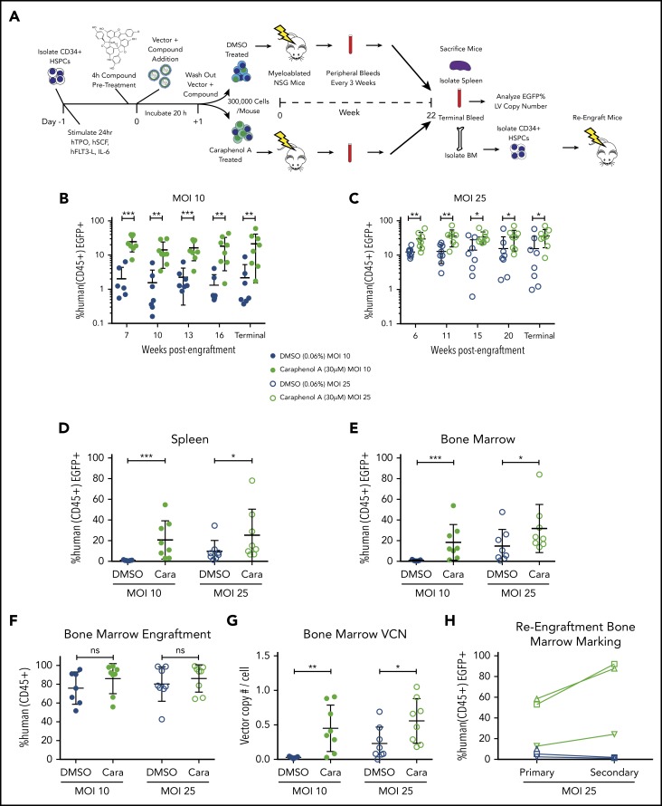
Figure 2.
Caraphenol A improves gene delivery to human HSCs in mice. (A) Experimental set-up of mouse transplant experiments. NSG mice were irradiated with 2.40 Gy. UCB-CD34+ cells from a pool of donors were thawed and prestimulated for 24 hours before a 4-hour incubation of caraphenol A or DMSO (n = 8 mice per treatment and MOI, 32 mice total) and LV, MOI = 10 (MOI 10) or MOI = 25 (MOI 25). Incubation with UCB-CD34+ cells lasted 20 hours, after which, 3 × 10⁵ cells per mouse were injected retro-orbitally, and the remaining UCB-CD34+ cells were cultured ex vivo. Transgene expression was measured 7 and 14 days posttransduction. Peripheral blood samples were obtained and evaluated every 3 to 5 weeks after an initial 6- to 7-week engraftment period. Mice were euthanized at 22 weeks (terminal) and peripheral blood, bone marrows, and spleens were obtained. For re-engraftment studies, CD34+ cells were isolated from MOI 25 cohort bone marrows (n = 3 from each treatment group), and 1 × 10⁵ cells were injected into irradiated NSG mice. Re-engraftment and gene marking were determined after 12 weeks. (B) Percentage human CD45+ EGFP+ cells in peripheral blood of UCB-CD34+ cell-engrafted NSG mice transduced with LV at either MOI = 10, (C) or MOI = 25, treatments as per the legend, throughout indicated points during the study period. Human cells were gated from the total leukocyte population and analyzed for EGFP expression. Dot plots presented (mean ± SD) with the y-axis in log10 scale. *P < .028, **P < .0042, ***P < .0006 by 2-tailed Mann-Whitney U test. Percentage human CD45+ EGFP+ cells in spleen (D) and bone marrow (E) of UCB-CD34+ cell engrafted NSG mice at the terminal time, comparing EGFP+ expression in caraphenol A- (green circles) and DMSO-treated (blue circles) mice at 2 MOIs. Dot plots presented (mean ± SD) comparing spleen MOI 10 (closed circles; ***P = .0003), MOI 25 (open circles; *P = .049) and bone marrow MOI 10 (closed circles; ***P = .0006), MOI 25 (open circles; *P = .042) by 2-tailed Mann-Whitney U test. (F) Comparison of donor engraftment in bone marrow at terminal time, as measured by total proportion of leukocytes that were mCD45− hCD45+. Dot plots presented (mean ± SD), n.s., not significant. (G) VCN of human cells from bone marrow of caraphenol A and DMSO-treated cohorts, 22 weeks after ex vivo LV transduction and compound treatment. VCN was recorded as a ratio of integrated Gag sequences per RNase P sequence. Dot plots presented (mean ± SD), comparing caraphenol A- with DMSO-treated mice at MOI 10 (closed circles; **P = .0022), and MOI 25 (open circles; *P = .022) by 2-tailed Mann-Whitney U test. (H) Percentage human CD45+ EGFP+ cells in bone marrow of NSG mice receiving UCB-CD34+ cells at terminal points of primary (left, 22 weeks) and secondary (right, 12 weeks) transplant, comparing EGFP+ expression in caraphenol A and DMSO mice at MOI 25. Data presented as dot plots, each representing individual mice and change from primary to secondary transplant.

