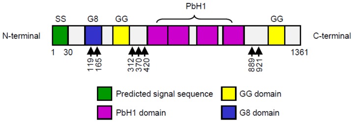
Figure 1.
Structure of full-length HYBID (hyaluronan binding protein involved in hyaluronan depolymerization, also known as KIAA1199). Functional domains of HYBID are indicated as follows: SS (signal sequence), predicted N-terminal signal sequence; G8, G8 domain; GG, GG domain; PbH1 (parallel β-helix repeats), PbH1 domain. The predicted N-glycosylation sites are shown by arrows. Numbers indicate the positions of the residues relative to the N-terminus of the full-length HYBID.

