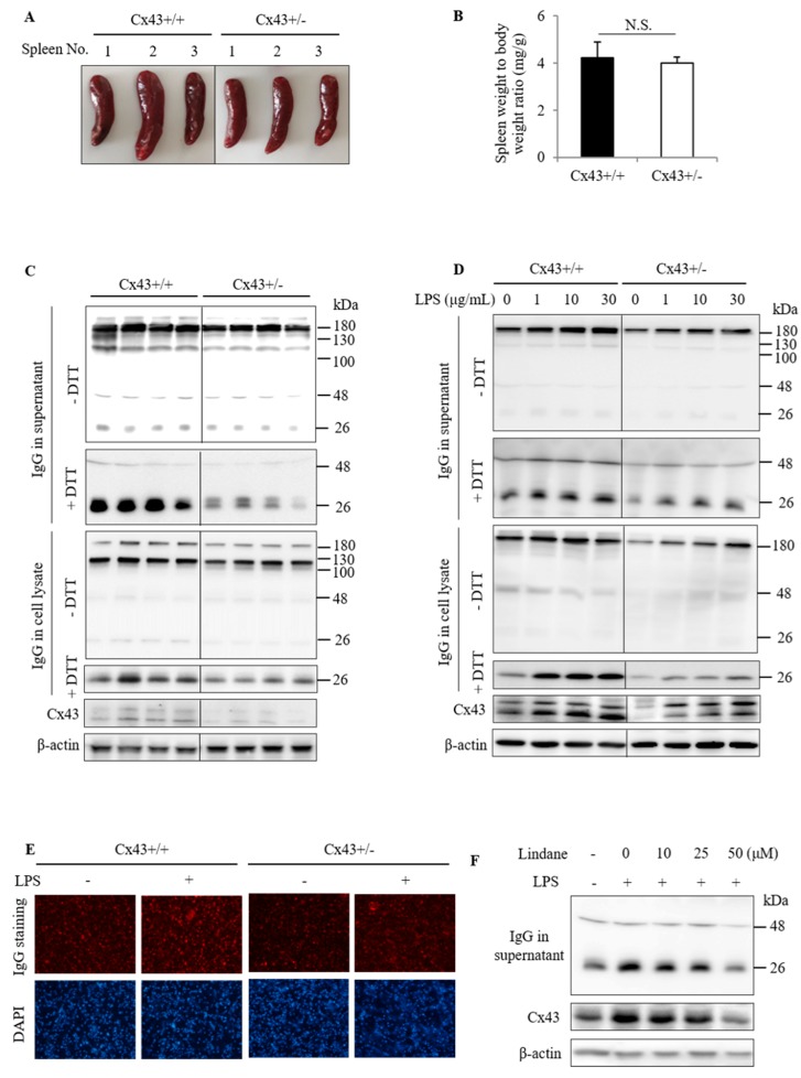
Figure 3.
Influence of Cx43 on spleen size and spleen cell production of IgG. (A,B) Spleen size and weight in Cx43+/+ and Cx43+/− mice. Spleen obtained from Cx43+/+ and Cx43+/− mice was photographed (A). The ratio of the spleen to bodyweight was shown in (B). Data are mean ± SE (n = 10). (C,D) IgG production by spleen cells from Cx43+/+ and Cx43+/− mice under basal and lipopolysaccharide (LPS)-stimulated condition. The spleen cells isolated from Cx43+/+ and Cx43+/− mice were cultured either for 2 days at basal condition (C) or in the presence of the indicated concentrations of LPS for 24 h (D). Supernatants and cell lysates were collected and analyzed for IgG by Western blot under native and denatured conditions. Protein level of Cx43 in the lysates was also detected. β-actin was used as an internal control. (E) Immunofluorescence staining of IgG in spleen cells from Cx43+/+ and Cx43+/− mice. The spleen cells were either left untreated or incubated with 20 μg/mL LPS for 16 h, and then subjected to immunofluorescence staining of IgG (red) and nuclear staining with DAPI (blue; magnification 200×). Note the different fluorescent intensity in Cx43+/+ and Cx43+/− spleen cells. (F) Effect of Cx43 inhibitor on LPS-induced IgG secretion. The spleen cells were stimulated with 20 μg/mL LPS in the presence or absence of the indicated concentrations of Lindane for 24 h. The collected supernatant and cell lysate were subjected to Western blot analysis for IgG and Cx43.

