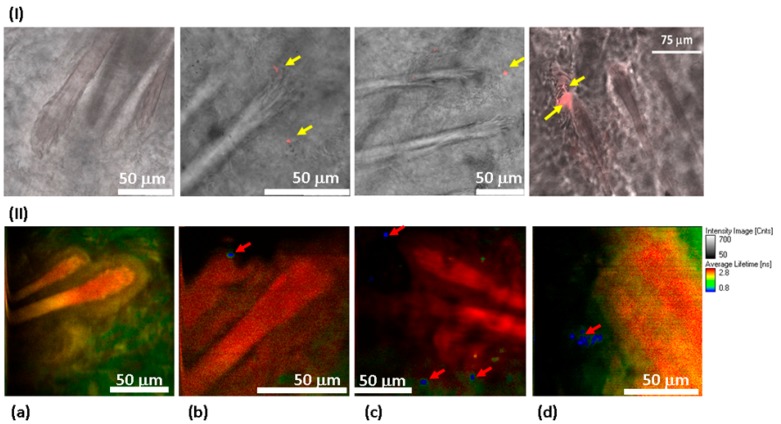
Figure 6.
Distribution of 100 ND in the skin using confocal imaging (I) and FLIM (II). 40× objective lenses (oil immersion for confocal measurements) were used. In confocal imaging (I), the fluorescence of 100 NDs was due to excitation at a 532 nm wavelength and was detected in the 650–720 nm range. In FLIM (II), a two-photon fluorescence was excited with an 800 nm femtosecond laser, and the signal was detected in the spectral range of 450–650 nm. (I) Skin sections observed using a confocal scanning microscope to visualize the fluorescence of 100 ND. No fluorescence signal was detected in the control sample (I(a)), while the treated sample presents a clear signal corresponding to the presence of 100 ND in different compartments of the hair follicles (shown in red and marked with yellow arrows) (I(b–d)). (II) FLIM: (II(a)) control sample. No fluorescence which could be related to 100 ND is detected. (II(b–d)) 100 ND-treated samples. The short lifetime signal (shown in blue) is visualized, which is characteristic of 100 ND.

