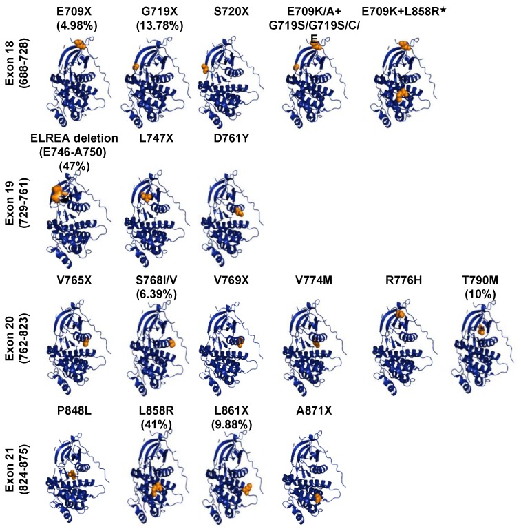
Figure 4.
EGFR mutations of kinase domain in NSCLC. Crystal structures of the kinase domain of EGFR are shown. Structures were drown using the PyMole Molecular Graphics System based on the protein Data Bank accession code 4R3P. The EGFR most frequent mutations in NSCLC, highlighted in orange, were mapped on the crystal structure of the EGF receptor’s kinase domain. The frequency of mutations are based on the COSMIC database of somatic mutations.

