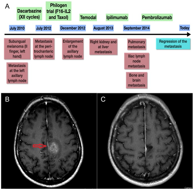
Figure 1.
Patient history. (A) Clinical history schematic of a representative patient who exhibited a loss of MSH6 expression in primary subungual melanoma tissue and in ileal and brain metastasis tissue. (B) CT scan of the patient's brain, presenting metastasis that was present prior to PD-1 treatment. (C) Last CT scan of the patient, presenting regression of the metastasis (red arrow) after treatment with anti PD-1. Green boxes represent the drugs administered, blue boxes represent follow-up times and red boxes denote the associated clinical-instrumental status. MSH6, clone 44 Ventana; PD-1, programmed death ligand 1.

