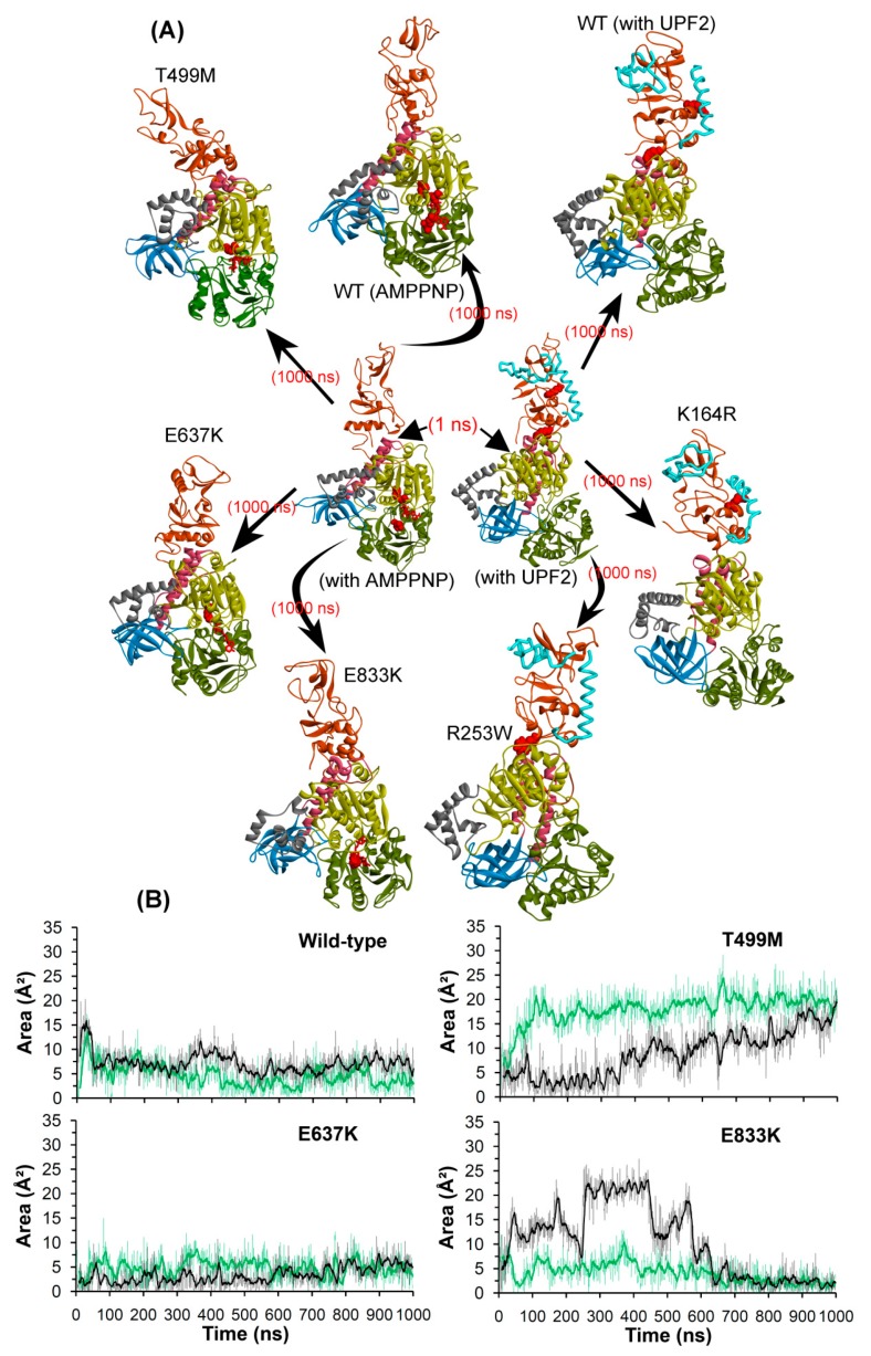
Figure 8.
Structural/conformational changes of UPF1 protein when complexed with UPF2 or AMPPNP. (A) Wild-type and mutant UPF1 protein in complex with AMPPNP or UPF2. (B) Area analysis of structural changes in the ATP-binding region of the UPF1 protein, triangle selected for area calculation was based on Cα atoms coordinates of T499, E637, and E833 residues. The dark lines represent the trend with a moving average of area with a period of 10 ns. Different domains of UPF1 are colored as per Figure 1.

