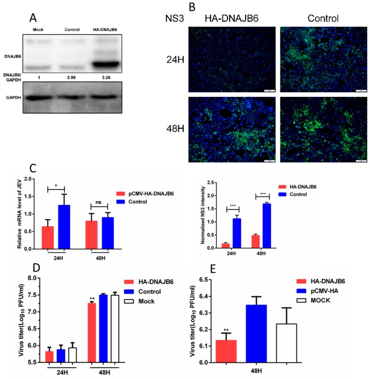
Figure 3.
Overexpression of DNAJB6 inhibits JEV infection. (A) Western blot analysis of HEK293 cells overexpressing DNAJB6. The relative DNAJB6 levels from the cells before they used for subsequent experiments were all determined using Western blot analysis to ensure DNAJB6 was overexpressed. The mock (untransfected) and control (transfected with empty vector) lanes illustrate the level of endogenous expression of DNAJB6. (B–D) Infection assays of HEK293 cells overexpressing DNAJB6 then infected with JEV at MOI of 1.0 for 24 h and/or 48 h. (B) JEV infection measured by NS3 protein (green) immunofluorescence. Scale bar, 100 µm. Quantitation of the JEV NS3 signal integrated density was normalized to the control cells (Mean ± SD, n = 3, Student’s t test; *** p < 0.001). (C) Viral mRNA levels measured by qRT-PCR (Mean ± SD, n = 3, Student’s t test; * p < 0.05, ns, not significant). (D) JEV titers measured by plaque assay (Mean ± SD, n = 3, one-way ANOVA; ** p < 0.01). (E) SK-N-SH cells overexpressing DNAJB6 then infected with JEV at MOI of 1.0 for 48 h. JEV titers were determined by plaque assay (Mean ± SD, n = 3, Student’s t test; ** p < 0.01).

